
What is Breast Thermography?

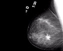
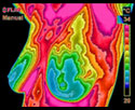
An infrared camera and computer system digital infrared imaging are used to measure heat from the surface of the breast to produce an image which can be evaluated for abnormalities. This modality has been in use since the 1960s and approved by the FDA in 1982 for use as an adjunctive breast cancer screening procedure. According to the American Cancer Society Breast Cancer Guidelines and Statistics you should have an initial infrared scan by age 20. Then a scan every 3 years between 20-30 years. At 30, you should have a scan every year.

The use of Digital Infrared Imaging (DII) is based on the principle that metabolic activity and vascular circulation in both pre-cancerous tissue and the area surrounding a developing breast cancer is almost always higher than in normal breast tissue. In an ever-increasing need for nutrients, cancerous tumors increase circulation to their cells by holding open existing blood vessels, opening dormant vessels, and creating new ones (neoangiogenesis). This process frequently results in an increase in regional surface temperatures of the breast. DII uses ultra-sensitive medical infrared cameras and sophisticated computers to detect, analyze, and produce high-resolution images of these temperature variations. Because of DII’s extreme sensitivity, these temperature variations may be among the earliest signs of breast cancer and/or a pre-cancerous state of the breast.

  • Gautherie, Ph.D.; Thermobiological Assessment of Benign and Malignant Breast Diseases. Am. J. Obstet. Gynecol., 1983; V 147, No. 8: 861-869.
  • Gamigami, M.D.; Atlas of Mammography: New Early Signs in Breast Cancer. Blackwell Science, 1996.
  • Keyserlingk, M.D.; Time to Reassess the Value of Infrared Breast Imaging? Oncology News Int., 1997; V 6, No. 9.
  • Ahlgren, M.D., E. Yu, M.D., J. Keyserlingk, M.D.; Is it Time to Reassess the Value of Infrared Breast Imaging? Primary Care & Cancer (NCI), 1998; V 18, No. 2.
  • Belliveau, M.D., J. Keyserlingk, M.D. et al ; Infrared Imaging of the Breast: Initial Reappraisal Using High-Resolution Digital Technology in 100 Successive Cases of Stage I and II Breast Cancer. Breast Journal, 1998; V 4, No. 4

Mammography

  • Uses X-rays to produce an image that is a shadow of dense structures. Suspicious areas need to be dense enough to be seen.
  • Structural imaging. Ability to locate the area of suspicious tissue.
  • Early detection method.
  • Findings increase suspicion. Cannot diagnose cancer.

    A biopsy is the only test that can determine if a suspected tissue area is cancerous.

  • Can detect tumors in the pre-invasive stage.
  • Average 80% Sensitivity (20% of cancers missed), in women over age 50. Sensitivity drops to 60% (40% of cancers missed) in women under age 50.
  • Hormone use decreases sensitivity.
  • Large, dense, and fibrocystic breasts cause reading difficulties.
  • In most women, the medial upper triangle, peripheral areas next to the chest wall, and the inframammary sulcus cannot be visualized.

Thermography

  • Uses infrared sensors to detect heat and increased vascularity (angiogenesis) as the byproduct of biochemical reactions. The heat is compiled into an image for computerized analysis.
  • Functional imaging. Detects physiologic changes. Cannot locate the exact area of suspicion inside the breast.
  • Early detection method. Used as an adjunctive imaging test.
  • Findings increase suspicion. Cannot diagnose cancer.

    A biopsy is the only test that can determine if a suspected tissue area is cancerous.

  • May provide the first signal that a problem is developing.
  • A positive infrared image represents the highest known risk factor for the existence of or future development of breast cancer – 10 times more significant than any family history of the disease.
  • Average 90% Sensitivity (10% of cancers missed) in all age groups.
  • No known effect.
  • No effect.
  • Not applicable.

Ultrasound

  • High frequency sound waves are bounced off the breast tissue and collected as an echo to produce an image.
  • Structural imaging. Ability to locate the area of suspicious tissue.
  • Low spatial resolution (cannot see fine detail). Good at distinguishing solid masses from fluid filled cysts. Used as an adjunctive imaging test.
  • Findings increase suspicion. Cannot diagnose cancer.

    A biopsy is the only test that can determine if a suspected tissue area is cancerous.

  • Ability to detect some cancers missed by mammography.
  • Average 83% Sensitivity (17% of cancers missed) in all age groups.
  • No known effect.
  • No known effect.
  • All areas visualized.

Sources:
Index Medicus – ACS, NEJM, JNCI, J Breast, J Radiology, J Clin Ultrasound
Index Medicus – Cancer, AJOG, Thermology
Text – Atlas of Mammography: New Early Signs in Breast Cancer
Text – Biomedical Thermology

This table “Mammography, Thermography and Ultrasound” is courtesy of Dr. William C. Amalu, DC, DABCT http://www.breastthermography.com/mammography_thermography.htm